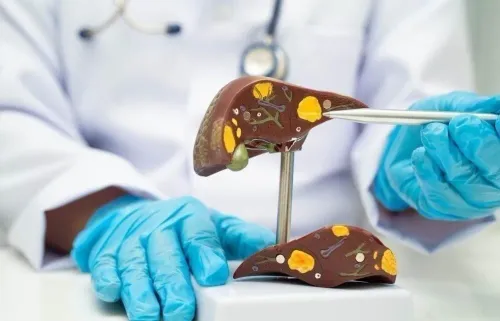

ERCP is not simply another scan: it uses a side-viewing endoscope and fluoroscopy to treat blockage, while MRCP and EUS usually provide diagnosis without entering the bile duct. By comparing how each route works, its risks, and the findings that justify it, you can ask more precise questions about the safest way to investigate or drain a bile duct problem.
Key takeaways
- ERCP treats bile duct problems; MRCP and EUS mainly investigate them.
- Choose ERCP when a blockage needs immediate treatment, not just imaging.
- Ask why surgery or percutaneous drainage is safer for your anatomy or condition.
- Before discharge, confirm the stent type, removal plan and warning symptoms.
What happens during ERCP, and when does treatment justify its risks?
ERCP is mainly a treatment procedure, not a routine diagnostic scan. A stomach doctor explaining ERCP for procedure comparison will describe a side-viewing duodenoscope passing through your mouth and stomach into the duodenum. The doctor identifies the papilla, injects contrast, and uses fluoroscopy to outline the bile and pancreatic ducts.
During the same session, the doctor can:
- Cut the sphincter (sphincterotomy) to open the duct.
- Remove stones with a balloon or basket.
- Widen a narrowed duct with dilation.
- Insert a plastic or metal stent to restore drainage.
- Take brushings or a biopsy for tissue sampling.
Choosing ERCP is most defensible when treatment is needed, such as for:
- A common bile duct stone causing obstruction or obstructive jaundice.
- Cholangitis requiring urgent drainage.
- A bile leak after surgery or injury.
- A bile duct stricture.
- Selected pancreatic duct blockages or leaks.
| ERCP | Uses fluoroscopy and duct access | Treats obstruction during the procedure |
|---|---|---|
| MRCP | Uses MRI without duct instrumentation | Maps ducts when diagnosis is uncertain |
| EUS | Views ducts from the stomach or duodenum | Finds small stones or masses; does not itself drain a duct |
If the only goal is anatomical diagnosis, MRCP or EUS usually carries less risk. ERCP can cause pancreatitis, bleeding, infection, perforation, or sedation complications without treatment benefit.
How do MRCP and EUS compare with ERCP?
MRCP and EUS are the main ERCP alternatives when the diagnosis is uncertain. MRCP uses MRI-based images to map the bile and pancreatic ducts without inserting instruments into the ducts, injecting contrast through the papilla, removing stones, or placing stents. It usually needs no sedation and gives a broad anatomical view.
EUS places an ultrasound probe in the stomach or duodenum to examine the nearby bile duct. It is particularly useful for small distal common bile duct stones, masses, and subtle duct abnormalities that other scans can miss. EUS is often more sensitive than MRCP for tiny stones, although both tests have high diagnostic accuracy.
| Feature | MRCP | EUS | ERCP |
|---|---|---|---|
| Purpose | Maps ducts for diagnosis | Finds small stones, masses, and subtle abnormalities | Treats an obstruction |
| Access | MRI scanner; no duct instrumentation | Probe through the mouth into the stomach or duodenum | Side-viewing scope reaches the papilla |
| Treatment | Cannot remove stones or place stents | Diagnostic EUS does not drain a duct | Can extract stones, dilate strictures, or place stents |
| Next step | ERCP if treatment is needed | ERCP can sometimes follow immediately | Treatment occurs during the procedure |
A negative MRCP or EUS does not exclude every tiny stone when pain, raised bilirubin, or a dilated duct keeps suspicion high. If you are searching for a stomach doctor ercp for procedure comparison, ask whether you need an anatomical diagnosis or immediate drainage; ERCP carries procedural risks and is usually reserved for treatment.
When are surgery or percutaneous drainage better bile duct procedure options?
Surgery or percutaneous drainage is better when the stone can be managed during gallbladder surgery, or ERCP cannot reach the papilla. These are important ERCP alternatives and bile duct procedure options.
Laparoscopic cholecystectomy removes the gallbladder; ERCP reaches an obstructed bile duct. If a duct stone appears during gallbladder surgery, the team may use ERCP before or after surgery, laparoscopic common bile duct exploration, or an intraoperative cholangiogram followed by treatment.
The choice depends on stone size and location, surgical expertise, pancreatitis or cholangitis, and altered anatomy.
| Option | What it does | When it applies |
|---|---|---|
| Laparoscopic cholecystectomy | Removes the gallbladder | Gallbladder stones require surgery |
| Common bile duct exploration | Removes duct stones during surgery | The surgeon has suitable expertise and access |
| ERCP | Extracts stones or places a stent through the papilla | The duct is reachable and needs treatment |
| PTBD | Drains bile through the skin and liver | ERCP cannot reach or cannulate the papilla |
| EUS-guided drainage | Creates internal drainage under ultrasound guidance | Failed ERCP in an expert center |
PTBD is useful when a tumor, prior surgery, or failed cannulation blocks ERCP access. It often leaves an external catheter and carries risks of bleeding, infection, bile leak, and catheter dislodgement.
EUS-guided biliary drainage may avoid that external catheter, but it is technically demanding. It is an expert-center rescue treatment after failed ERCP, not an ordinary diagnostic EUS examination.
How do doctors decide whether choosing ERCP is justified now?
Direct ERCP is justified now when the evidence shows an obstructed bile duct that needs treatment, not merely a duct that needs mapping.
Doctors combine the suspected cause with ultrasound or CT findings, bilirubin and liver-enzyme results, common bile duct diameter, fever or sepsis, bleeding risk, previous surgery, and whether you need drainage immediately or imaging only.
For suspected common bile duct stones, the practical risk framework is:
| Finding | Usual implication | Next step |
|---|---|---|
| Stone visible on ultrasound or CT | High risk of obstruction | Direct ERCP for removal or drainage |
| Acute cholangitis | Infected, obstructed bile duct | Antibiotics, resuscitation, and urgent drainage |
| Markedly abnormal bilirubin plus a dilated common bile duct | High likelihood of a stone | Direct ERCP |
| Abnormal tests without high-risk imaging or infection | Intermediate risk | EUS, MRCP, or intraoperative cholangiography |
| Altered anatomy or high bleeding risk | Access or complication concerns | Adapt the bile duct procedure options and consider specialist alternatives |
Cholangitis needs particular urgency: antibiotics and supportive care do not replace drainage. When feasible, endoscopic drainage is preferred over percutaneous drainage, with decompression targeted within 48 hours and earlier treatment for severe or deteriorating illness.
Previous surgery can make papilla access difficult, while anticoagulants, low platelets, or clotting problems affect sphincterotomy planning. For a consultation with Dr Ravindra Surude 08042756059 in Mira Road East, bring scan reports, bilirubin and liver-test results, medication details, prior operative records, and a clear question about choosing ERCP for drainage versus diagnosis.
Related services
Gastrointestinal endoscopy procedures ERCP ( Endoscopic Retrograde cholangiopancreatography ) ERCP (Endoscopic Retrograde Cholangiopancreatography) by Dr. Ravindra Surude :Expert Diagnosis and Treatment of Pancreatobiliary Disorder Dr. View service → |
Gastrointestinal Diseases Pancreatobiliary Disorders Treatments Pancreatobiliary Disorder Treatment by Dr. Ravindra Surude Expert in Pancreas, Bile Duct & Gallbladder Diseases | Mumbai & Thane Dr. View service → |
What should you confirm before ERCP and before leaving with a stent?
Before choosing ERCP, confirm that treatment is worth its practical burden. It usually involves deep sedation or general anesthesia, fluoroscopy, and recovery restrictions such as no driving, alcohol, or important decisions until the sedation has cleared.
Post-ERCP pancreatitis is the most common serious complication, so compare this risk with ERCP alternatives such as MRCP or EUS.
| Question | What to confirm | Why it matters |
|---|---|---|
| Risk reduction | Is rectal indomethacin or diclofenac suitable? Will wire-guided cannulation be used? Is a prophylactic pancreatic-duct stent needed for this high-risk case? | These measures reduce pancreatitis risk but do not remove it. |
| Stent plan | Is the stent plastic or metal, and where will it sit? | Plastic stents generally need scheduled removal or exchange. |
| Follow-up | What is the intended duration, and what is the appointment date? | An occluded or migrated stent can cause recurrent jaundice or cholangitis. |
Before discharge, request the stent type, location, intended duration, and follow-up appointment in writing. Keep the report with your medication list and seek urgent care for fever, worsening abdominal pain, vomiting, or yellowing of the eyes.
For persistent or malignant obstruction, ERCP can drain the duct and sometimes obtain tissue, but it does not cure cancer. Uncovered versus covered metal stents, drainage of one or both liver systems, surgery, and oncology treatment depend on anatomy, resectability, expected survival, and the overall treatment plan.
Frequently asked questions
What happens during ERCP, and when does treatment justify its risks?
A doctor passes a side-viewing duodenoscope through the mouth to the duodenum, injects contrast, and uses fluoroscopy to examine the ducts. Treatment can include sphincterotomy, stone removal, dilation or stent placement. Its risks are easier to justify when a confirmed blockage, infected bile duct or bile leak needs treatment during the same procedure.
How do MRCP and EUS compare with ERCP?
MRCP is a non-invasive MRI scan that maps the bile and pancreatic ducts without inserting an endoscope. EUS uses an ultrasound probe on an endoscope to examine the bile duct, gallbladder, pancreas and nearby lymph nodes, and can obtain tissue samples. ERCP is more invasive but allows treatment during the procedure.
When are surgery or percutaneous drainage better bile duct procedure options?
Surgery can be preferable when a problem needs an operation, such as gallbladder removal or reconstruction, or when endoscopic access is unlikely to work. Percutaneous transhepatic drainage can provide access through the skin and liver when ERCP cannot reach the obstruction or has failed.
How do doctors decide whether choosing ERCP is justified now?
They consider the suspected diagnosis, imaging findings, symptoms, infection, liver test results, anatomy, previous procedures, bleeding risk and whether treatment is needed immediately. A diagnostic MRCP or EUS is often more appropriate when no intervention is planned.
What should you confirm before ERCP and before leaving with a stent?
Confirm the purpose of ERCP, planned treatment, sedation or anaesthesia, medication instructions, fasting requirements and alternatives. Before discharge, ask for the stent type and location, the removal or exchange date, follow-up arrangements and urgent symptoms such as fever, severe abdominal pain, vomiting or black stools.
Related service
Gastrointestinal endoscopy procedures Endoscopic Ultrasound (EUS) by Dr. Ravindra Surude Advanced Diagnostic & Therapeutic GI Imaging Dr. View service → |